
Ultrasound
MicroFlow (MFI) and MicroFlow-HD (MFI-HD) imaging mode
Detect low-velocity micro-vessel anatomy in tissue without using contrast
MicroFlow Imaging (MFI) detects slow and weak blood flow anatomy in tissue. It maintains a high frame rate and excellent 2D image quality while applying artifact reduction techniques. 2D image subtraction, 2D blending and side-by-side display options offer versatility in visualization.
MFI-HD offers twice the sensitivity and resolution of MFI in assessing blood flow1. MFI HD is well-suited for studies requiring high resolution and sensitivity, including renal/abdominal, breast, MSK, small parts, CEUS, and OB/GYN exams.
Spatial-temporal clutter filter
MFI’s novel spatial-temporal filter analyzes Doppler data, evaluating signal characteristics from both a spatial and temporal dimension in order to distinguish the flow signals from the tissue clutter signals. In this manner, MFI can detect the lowest velocity blood flow, which microvascular vessels typically exhibit, and remove distracting tissue clutter.

Vessel enhancement algorithm
The vessel enhancement algorithm in MFI-HD reduces noise and enhances detected vessels1 without introducing blurring often seen with more traditional image processing techniques. This vessel enhancement combines with acoustic transmits designed to optimize flow sensitivity and spatial resolution to improve visualization with high spatial resolution.

Four display modes
MFI HD features four display modes based on if tissue subtraction and echo-flow compare are used, allowing customization of images. When tissue subtraction is on, MFI is focuses on displaying only the flow information, and therefore this provides the most sensitive display method. When tissue subtraction is off, MFI utilizes a tissue-flow arbitration algorithm to seamlessly blend flow with tissue information in a smooth and natural manner, therefore providing anatomical context to the visualized flow. It has the additional benefit of further tissue artifact suppression.

Image gallery
- Kidney
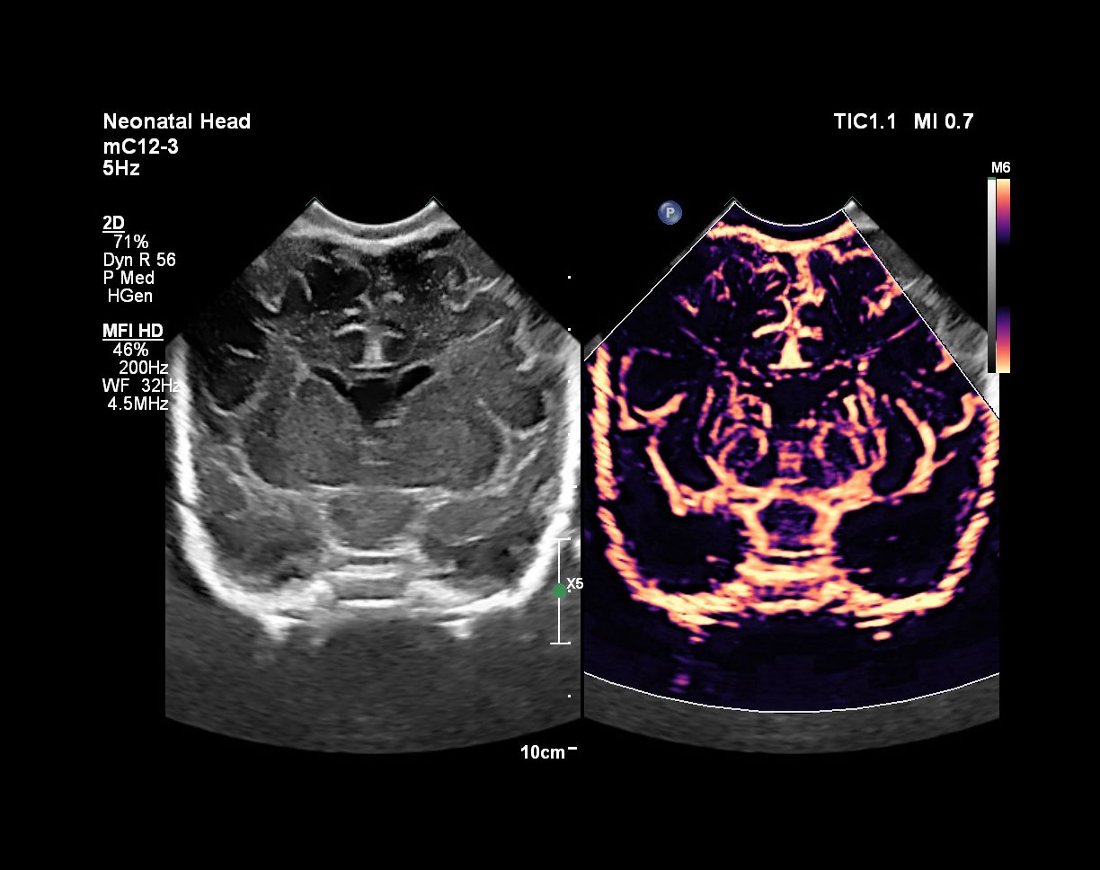
- Neonatal Head
- Breast Cysts
- Contrast Enhanced Ultrasound(CEUS) with Micro Flow Imaging(MFI) of a Focal Nodular Hyperplasia
- Portal Vein
- Superficial Thumb
- Superficial Finger
- Neonatal Head
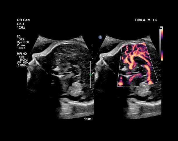
- Pulmonary vein and fetal heart MFI with Flow Viewer using C5-1 transducer
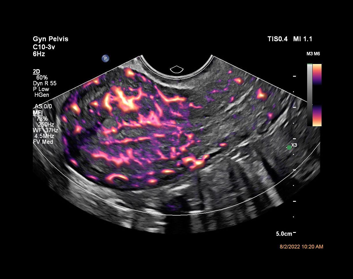
- Ovarian blood flow with MFI using C10-3v transducer
- Fetal brain pericallosal artery with MFI HD using C5-1 transducer
- Fetal lung perfusion with MFI HD using eL18-4 transducer
- Fetal circle of Willis with MFI HD
- Fetal ductus venous with MFI HD
- Flow Viewer applied to MFI with C10-3v uterus


mL26-8 compact and ultra-high frequency transducer
Philips mL26-8 ultra-high frequency, micro-linear transducer allows you to image from eyeball to hip - all with the same, fully versatile transducer. Added presets for MSK, breast, vascular, dermal and ocular provide added versatility.

Footnotes
- Internal measured comparison on standards MFI to MFI HD using clinical targets and standard measurement methodology. Not available on Affiniti systems.
Disclaimer
*Based on a sample size of 21 users.
**Based on a sample size of 20 users.